How Do They X Ray Baby Hips
As the hips are moved in these tests a hip click can be felt by the examiner. Around 6 months of age enough bone is present in an infant hip to make an X-ray more accurate than ultrasound.
 Ortho Dx A Woman With A Hip That Is Popping Out Of Place Clinical Advisor
Hip X-rays pass through skin and soft tissue mostly but do not pass through bone or metal easily.
How do they x ray baby hips. Second view of the hip. Most children do not need surgery but for those who do an arthrogram x-ray dye injected into the hip joint at the beginning of the surgery can help the surgeon decide exactly what needs to be corrected. In addition a baby will be sent for ultrasound if the doctor finds an abnormality of the hip during a physical examination such as.
Ultrasounds are the diagnostic method of choice for infants under 6 months of age. When should I order an X-ray rather than an ultrasound to diagnose a musculoskeletal problem in an infant. The lumbar spine is made up of five vertebral bones.
It turns out this photo originally posted on Reddit of a baby squished into a tube like a lil baby deposit is actually of him getting a tiny X-ray. An X-ray procedure may take just a few minutes for a simple X-ray or longer for more-involved procedures such as those using a contrast medium. Shoot through lateral with the good leg flexed.
The sacrum is. The examination involves gently moving your babys hip joints to check if there are any problems. I would also recommend swimming a dog to build muscle mass if there is any question on the hips.
This lets the radiologist to get X-ray pictures. X-rays are usually carried out in hospital X-ray departments by trained specialists called radiographers although they can also be done by other healthcare professionals such as dentists. As different tissues in the body absorb different amounts of radiation the images will show different shades of black and white.
X-rays have more energy than rays of visible light or radio waves. A hip X-ray is a safe and painless test that uses a small amount of radiation to make images of the hip joints where the legs attach to the pelvis. You will go in the room with him he will need to be stripped from the waist down they will take x-rays of him flat on his back legs dead straight and together you wil be able to hold him in this position then an x-ray of his still on his back with his knees bent facing outwards and the soles of his feet put together he will be fine its not traumatic at all you will be with him the whole time.
The better condition a dog is in the better chance of a good x-ray. Two tests are performed called the Barlow and Ortolani tests to examine the function of the hip joints. He or she may use pillows or sandbags to help you hold the position.
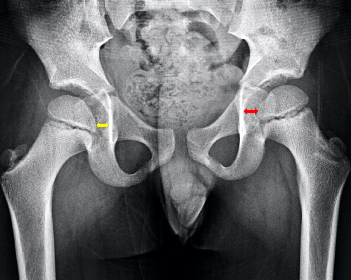
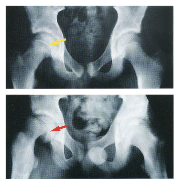
During the X-ray exposure you remain still and sometimes hold your breath to avoid moving so that the image doesnt blur. I have a friend who has watched the OFA on a yearly. This x-ray shows a dislocated hip on the patients right side.
The contraption hes in is called a Pigg O Stat. During the examination an X-ray machine sends a beam of radiation through the pelvic bones and hip joints and an image is recorded on a computer or special film. You can then view these pictures on a photographic film or on a computer monitor.
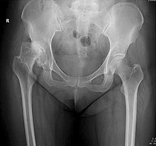
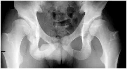
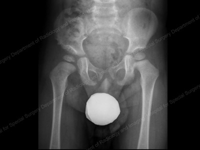
Its a cast that goes around both hips and down the leg to keep the hips aligned. AP radiograph of the pelvis. An X-ray of the pelvis focuses specifically on the area between your hips that holds many of your reproductive and digestive organs.
Patients left hip is dislocated. The Pavlik harness is used on babies up to four months old to hold their hip in place while allowing their legs some movement. So I redid the x-ray at 9 months and saw an entirely different x-ray.
Your pelvis is made up of three bones the ilium ischium and. The dog will pass OFA if the x-ray stays the same. X-rays are forms of radiant energy like light or radio waves.
A lumbosacral spine X-ray or lumbar spine X-ray is an imaging test that helps your doctor view the anatomy of your lower back. Used for general pelvis assessment eg. Im a nurse in a pedi operating room and Ive done them many times The hip dysplasia usually resolves on its own with the casting or bracing as the baby gets older.
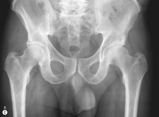
Just the pelvis x-ray. With early diagnosis and treatment most children are able to develop normally and have a full range of movement in their hip. How to administer APLat x-rays of the pelvis and hips for basic x-ray operators.
It is put on by an orthopedic surgeon while using x-ray to make sure the hip is aligned correctly. X-rays are a type of radiation that can pass through the body. They can penetrate your body.
Typically this takes about eight to 12 weeks. They cant be seen by the naked eye and you cant feel them. The baby usually wears the harness all day and night until their hip is stable and an ultrasound shows their hip is developing normally.
Your babys hips will be checked as part of the newborn physical examination within 72 hours of being born. In babies with hip dysplasia the joint has not formed normally and the hips are prone to moving in and out of joint. At birth an inability to move the thigh outward at the hip as far as normally possible a hip click heard or felt by the doctor when moving the infants thigh.
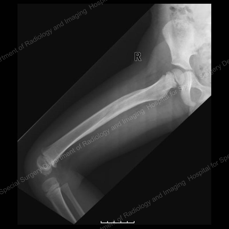 Femur Fractures In Children Treating A Child S Broken Thighbone
X Ray Of Hip Dysplasia Wikipedia
 Pelvis And Hips Radiology Key
 Pediatric Hip Disorders Radsource
 Developmental Dysplasia Of Hip Or Congenital Dislocation Of Hip Bone And Spine Radiology Developmental Dysplasia Of The Hip Diagnostic Imaging
 Interpreting X Rays Of The Pelvis Hip Joint And Femur Youtube
 Hip Dysplasia Information Symptoms Diagnosis Treatment
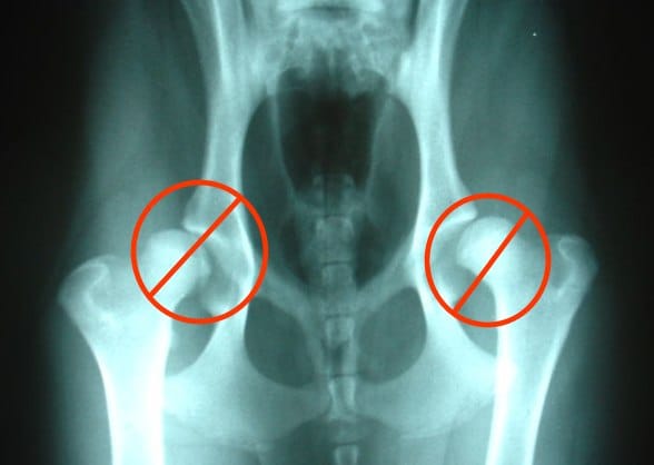
 Leerburg The Importance Of Good Positioning On Canine Hip X Rays
 Congenital Hip Dysplasia Symptoms Treatments Orthopedics
 Hip Dysplasia Adolescent Description
 Pediatric Hip Frog Leg Lateral View Radiology Reference Article Radiopaedia Org
 The Radiology Assistant Hip Pathology In Children
 Hip Dysplasia In Dogs Part 2 The Real Cost Of Diagnosis Petmd
 Leg Lengths After Surgery Total Hip Surgery Pagets Disease Avascular Necrosis Radiology
 Developmental Dysplasia Of The Hip Radiology Reference Article Radiopaedia Org
 Hip Elbow Information
 Developmental Hip Dysplasia In Babies And Young Children
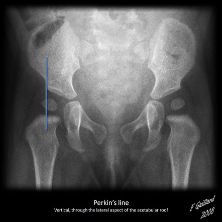
 Diagnosis International Hip Dysplasia Institute
 Slipped Capital Femoral Epiphysis Scfe
Post a Comment for "How Do They X Ray Baby Hips"